Microorganisms of Food Safety Concern
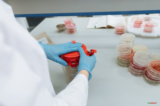
Better understanding of the causative agents of spoilage and poisoning in food is the foundation to food safety. Here are the most common bacteria and parasites of food safety concern
1. Staphylococcus aureus (Staphylococcosis)
Staphylococcus aureus is gram positive cocci that occur in singles, short chains and irregular grape like cluster. Only the strains that produce enterotoxin can cause food poisoning.
Epidemiology
The most important sources to foods are nasal carriers and individuals whose hands and arms are inflicted with boils and sores, who are permitted to handle foods (Quinn, Markey, Carter, Demelly, & Leonard, 2001).
Pathogenesis
If food is stored for some times in room temperature the organism may grow in the food and can produce toxin. The bacteria produce enterotoxin while multiplying in food. S. aureus is known to produce six serologically different types of enterotoxins (A, B, C1, C2, D and E) that differ in toxicity.
Most food poisoning is caused by enterotoxin A followed by type D. these enterotoxins are heat stable, with type B being most heat resistant. Enterotoxin stimulates the central Nervous Systems (CNS) vomiting and others, are classed as bacterial super antigens relative to in vivo antigen recognition in contrast to conventional antigens (Quinn, et al., 2013).
Symptoms
It is characterized by a short incubation period typically 2-4 hours. The onset is sudden and is characterized by vomiting and diarrhoea but no fever. The illness lasts less than 12 hours.
In severe cases dehydration, and collapse may require treatment through intravenously infusion. The short incubation periods are the characteristics of intoxication where illness in the results of ingestion of the preformed toxin in the food (Adams & Moss, 2008).
Detection of the organism
The presence of a large number of S. aureus organism in a food indicate poor handling or sanitation. The dilution is placed on baird-parker agar or mannitol salt agar. The enterotoxin can be detected and identified by gel diffusion (Quinn, Markey, Carter, Demelly, & Leonard, 2001; Radostits, Gay, Hinchlif, & Constable, 2007).
Prevention and control
Can be prevented and control by proper cooking and preparing food as well as storing.
- Control measures include education of those who prepare the food at home and other food handlers to take proper personal measures.
- Prohibiting individuals with sores or other skin lesions from handling food is the second intervention.
- Lastly, place food in cold place at 4°C or lower of all food in order to prevent bacterial multiplication and formation of toxin. Foods must be kept at room temperature for as little as possible (WHO, 2008).
The occurrence is worldwide but varies depending on conditions of food hygiene.
2. Vibrio cholerae (Cholera)
Vibrio cholerae 01 and 0139 are the bacterial agents for the disease. V. cholarae 01 includes two biotypes- classical and E1 Tor- each of which includes organisms of Ogawa, Inaba and rarely Hikojima serotypes.
Epidemiology
They are Gram-negative, facultative anaerobic, motile, non-spore forming rods that grow at 18-42°C (optimum 37°C), pH 6-11 (optimum 7.6) water activity of 0.97.
Growth is stimulated by salinity levels of around 3% but prevented by levels of 6%. Organism is resistant to freezing but sensitive to heat and acid (National Disease Surveillance Centre, 2004).
Pathogenesis
May survive for some days on fruit and vegetables. It is often found in aquatic environments and is part of the normal flora in brackish water and estuaries. V. cholera is non-invasive and diarrhoea is mediated by cholera toxin formed in the gut (toxico-infection).
Symptoms
Incubation period 1-3 days. Profuse watery diarrhoea, which can lead to severe dehydration, collapse and death within a few hours unless lost fluid and salt are replaced; abdominal pain and vomiting. Duration is up to 7 days.
Mode of transmission
Food and water contaminated through contact with faecal matter or infected food handlers. Contamination of vegetables may occur through sewage or wastewater used for irrigation.
Person to person transmission through the fecal-oral route is also an important mode of transmission. Foods involved include seafood, vegetables, cooked rice and ice.
Control and Prevention
Safe disposal of sewage and wastewater, treatment of drinking water e.g. chlorination or irradiation, heat treatment of foods e.g. canning; high pressure treatment; good hygiene practices during production and processing.
In food service establishment/household, personal hygiene (hand washing with soap and water); thorough cooking of food and careful washing of fruit and vegetables; boiling drinking-water when safe water is not available. Consumers should avoid eating raw seafood.
Occurrences
Africa, Asia, parts of Europe and Latin America. In most industrialized countries, reported cholera cases are imported by travellers or occurs as a result of imported food.
3. Listeria spp (Listeriosis)
Listeria monocytogenes is the bacterium that causes the infection listeriosis. It is a facultative anaerobic bacterium, capable of surviving in the presence of oxygen.
It can grow and reproduce inside the host’s cells and is one of the most virulent food-borne pathogens, with 20 to 30 percent of clinical infections resulting in death (van-de-Venter, 2000).
Epidemiology
Listeria species are widely distributed in the environment and can be isolated from soil, plants, decaying vegetation and silage (pH 5.5) in which the bacteria can multiply. Asymptomatic faecal carrier occurs in man and animal species (Ibid).
Pathogenesis
Listeria originally evolved to invade membranes of the intestines, as an intracellular infection, and developed a chemical mechanism to do so. This involves a bacterial protein “internalin” which attaches to a protein on the intestinal cell membrane “cadherin”.
L. monocytogenes has also D-Galactose residues on its surface that can attach to D-Galactose receptors on the host cell walls. These host cells are generally M cells and Payer’s patches of the intestinal mucosa.
Once attached to these cells, L. monocytogenes can translocate past the intestinal membrane and into the body. L. monocytogenes may invade the gastrointestinal epithelium. Once the bacterium enters the host’s monocytes, macrophages, or polymorphonuclear leukocytes, it becomes blood-borne (septicaemic) and can grow.
Its presence intracellularly in phagocytic cells also permits access to the brain and probably trans-placental migration to the foetus in pregnant women (CDC, 2011).
Symptoms
The symptoms of listeriosis usually last 7-10 days, with the most common symptoms being fever, muscle aches, and vomiting. Diarrhoea is another, but less common symptom. If the infection spreads to the nervous system it can cause meningitis, an infection of the covering of the brain and spinal cord (Bean & Griffins, 1990; Adams & Moss, 2008).
Detection of toxin
Enrichment procedures are required for this organism. This involves inoculation of selective or non-selective broths that are incubated at 4°C for up to 8 weeks.
An ELISA, using monoclonal antibodies, has been developed to identify listeria in food, and also DNA probe for detection of bacterium in dairy products (Niehaus, et al., 2011).
Control and prevention
The main means of prevention is through the promotion of safe handling, cooking and consumption of food. This includes washing raw vegetables and cooking raw food thoroughly as well as reheating leftover or ready-to-eat foods like hot dogs until steaming hot (CDC, 2011).
Preventing listeriosis as a food illness requires effective sanitation of food contact surfaces. Alcohol and Quaternary ammonium are an effective topical sanitizer against Listeria.
Refrigerated foods in the home should be kept below 4°C (39.2°F) to discourage bacterial growth. Preventing listeriosis also can be done by carrying out an effective sanitation of food contact surfaces.
Occurrence is worldwide. Most cases have been reported from Europe, North America and the Pacific islands.
4. Campylobacter spp (Campylobacteriosis)
Campylobacteriosis is an infection caused by bacteria of the genus Campylobacter. There are approximately sixteen species associated with Campylobacter, but the most commonly isolated are C. jejuni, C. coli, and C. upsaliensis.
The most prevalent species associated with human illness is C. jejuni. Campylobacter is also responsible for 15% of foodborne illness-related hospitalizations, and 6% of foodborne illness-related deaths (Hoffmann, et al., 2017).
Epidemiology
Campylobacter is one of the most common causes of human bacterial gastroenteritis. A large animal reservoir is present as well, with up to 100% of poultry, including chickens, turkeys, and waterfowl, having asymptomatic infections in their intestinal tracts.
Infected chicken feces may contain up to 10^9 bacteria per 25 grams, and due to the installations, the bacteria are rapidly spread to other chickens. This vastly exceeds the infectious dose of 1000-10,000 bacteria for human (Hoffmann, et al., 2017).
Pathogenesis
Bacterial motility, mucus colonization, toxin production, attachment, internalization, and translocation are among the processes associated with C. jejuni virulence. Infection begins with ingestion of the C. jejuni in contaminated foods or water.
Gastric acid provides a barrier, and the bacteria must reach the small and large intestines to multiply; C. jejuni invades both epithelial cells and cells within the lamina propria (Hoffmann, et al., 2017).
Symptoms
The symptoms associated with this disease are usually flu-like fever, nausea, abdominal cramping, vomiting, enteritis, diarrhoea, and malaise. Symptoms begin within 2-5 days after ingestion of the bacteria, and the illness typically lasts 7-10 days.
Recurrence of this disease can occur up to three months after pathogen ingestion (Shonhiwa, Ntshoe, Essel, Thomas, & McCarthy, 2018). Other complications can include meningitis, urinary tract infections and short-term reactive arthritis.
Detection of Toxin
Because of the unique growth characteristics of Campylobacter, isolation of these organisms from field samples requires the use of special media and culture conditions. Campylobacter jejuni and Campylobacter coli can be isolated from the intestines of healthy farm animals, poultry, pets, zoo animals, and wild birds.
Diagnosis of C. jejuni is based on isolation of the organism on selective media under micro aerophilic conditions. PCR-based methods are effective in identifying infection especially if cultivation is difficult or if the sample has been somewhat mishandled.
However, a positive test is not sufficient evidence to determine causation and must be considered in conjunction with clinical signs (van-de-Venter, 2000).
Control and Prevention
Control depends on sanitation and hygiene in livestock barns to reduce the bacterial populations in the environment of the animals. The number of organisms can be reduced and controlled in meat processing plants by using HACCP protocols including the washing, handling and freezing of carcasses.
Improvement of food-handling skills in restaurants and in the home, kitchen will reduce transmission of the organism and adequate cooking of raw meat such as poultry to an internal temperature of 82°C will eliminate the organism (Walderhaug, 2007).
Occurrence is worldwide. It is one of the most frequently reported foodborne diseases in industrialized countries; a major cause of infant and travellers’ diarrhoea in developing countries. Campylobacter spp cause 10-15% of cases of diarrhoeal diseases in children seen at treatment centres.
5. Shigella spp (Shigellosis)
Shigella is a species of enteric bacteria that causes disease in humans and other primates. Shigella is gram-negative rods that are non-motile and non-spore forming.
The bacteria are primarily a human disease but has been found in some primates. Shigella are facultative anaerobes, similar to enterics such as E. coli (Brayan, Mckiley, & Mixon, 1971).
Epidemiology
Shigella transmission can occur through direct person-to-person spread or from contaminated food and water. The minimal infectious dose can be transmitted directly from contaminated fingers, since intermediate bacterial replication is not required to achieve the low infectious dose.
In developed countries, most cases are transmitted by faecal-oral spread from people with symptomatic infection. In developing countries, both faecal-oral spread and contamination of common food and water supplies are important mechanisms of transmission symptom.
Pathogenesis
Shigella attaches to and penetrate intestinal cell walls of the small intestines by producing toxins that may promote the diarrhoea characteristic of the disease. The Shiga toxin enables the bacteria to penetrate the epithelial lining of the intestines, leading to a breakdown of the lining and haemorrhage.
Shigella also have adhesins that promote binding to epithelial cell surfaces and invasion plasmid antigens that allow the bacteria to enter target cells, thus increasing its virulence (CDC, 2011).
Symptoms
They include abdominal pain, cramps, diarrhoea, fever, vomiting, blood, pus or mucus in stools. Mild infections cause low-grade fever (38 – 38.9°C) and watery diarrhoea 1 to 2 days after people ingest the bacteria.
Abdominal cramps and a frequent urge to defecate are common with more severe infections. Children, particularly young children, are most likely to have severe complications of high fever (41°C) sometimes with delirium. Severe dehydration with weigh loss is also a symptom.
Detection of toxin
Shigella infection is diagnosed through testing of a stool sample. First a stool sample must be obtained from the potentially infected person, and then the sample is placed on a medium to encourage the growth of bacteria.
If and when there is growth, the bacteria are identified, usually by looking at the growth under a microscope (Ibid).
Control and prevention
Shigella is heat-sensitive and will be killed by thorough heating (over 70°C). Raw or undercooked foods and cross-contamination, when cooked material comes into contact with raw produce or contaminated materials (cutting boards), are the main causes of infection.
Proper cooking and hygienic food handling thus can prevent Shigella infections to a large extend. There is currently no vaccine for Shigellosis prevention, but there is current research that appears promising.
The most effective method for prevention is frequent and vigorous hand washing with warm, soapy water and ensuring clean drinking water sources and proper sewage disposal in developing nations (Malangu, 2016).
Occurrence is worldwide and has higher prevalence in developing countries. Shigellosis is a major cause of diarrhoea in infants and children under the age of 5 years and accounts for 5-15% of diarrhoeal diseases cases seen at treatment centres.
6. Escherichia coli
Escherichia coli, commonly known as E. coli, is a bacterium that resides in the intestines of humans and animals such as cows, sheep, and goats. While most strains are harmless and aid in digestion, certain types can cause serious illness, including food poisoning.
E. coli belongs to the Enterobacteriaceae family and is characterized as a gram-negative rod, up to 3 micrometers long. It ferments glucose and various sugars, producing pink colonies on McConkey agar. Certain strains exhibit hemolytic activity on blood agar and are motile with peritrichous flagella, often featuring fimbriae (hair-like structures). E. coli 0157:H7 is an important serotype and seems to be predominate in most areas. The strains producing verotoxin are shiga-like toxin (SLT) which causes diarrhoea in humans and animals.
Source of infection
Contamination of food by human and animal faeces. The organism can persist in manure, water trough and other farm location. The association of E. coli 0157:H7 with raw meat, under cooked ground beef and raw milk lead to investigation of the role of cattle as a reservoir of the pathogens (Buchanan & Doyle, 1997).
Pathogenesis
Enterohemorrhagic E. coli (EHEC) strain, particularly the serotype E. coli O157:H7, may produce one or more types of cytotoxins which are collectively referred as shiga-like toxins (SLTs) since they are antigenically and functionally similar to shiga toxin produced by Shigella dysenteriae.
However, new terminology has been applied, and what was SLT is now Stx. All Stxs consists of a single enzymatically active A subunits and multiple B subunits. Stx-sensitive cells possess the toxin receptor, globotriaosyceramide (Gb3), and sodium butyrate appears to play a role in sensitizing cells to Stxs.
Once toxins bind toGb3, internalization follows with transport to the trans-Golgi network. Inside the host cells, the A subunits bind to and release and adenine residue that inhibits protein synthesis. The B subunits form pentamers in association with a single A subunit and thus are responsible for the binding of the toxin to the neutral glycolipid receptors. E. coli O157:H7 thrives in diverse conditions (7°C to 50°C) and acidic environments but is sensitive to heat.
Symptoms
Infection with E. coli O157:H7 typically manifests after an incubation period of 72-120 hours (3-5 days). Symptoms include:
- Diarrhea: Initially watery, often progressing to bloody
- Abdominal cramps: Severe and persistent
- Vomiting: Occasional
- Absence of fever, unlike many bacterial infections
Most cases resolve within a week, but severe infections can lead to hemolytic uremic syndrome (HUS), a life-threatening condition affecting the kidneys, especially in children and the elderly. Seek medical attention immediately if bloody diarrhea or intense abdominal pain occurs. Antibiotics are generally avoided, as they may worsen HUS risk.
Sources and Transmission
E. coli spreads through:
- Contaminated food: Undercooked ground beef, raw meat, raw milk, or produce exposed to human or animal faeces
- Water: Drinking or swimming in contaminated sources
- Cross-contamination: Improper handling of raw and cooked foods
- Person-to-person: Poor hygiene after contact with infected individuals
- Animal contact: Interaction with farm animals or petting zoos
Cattle serve as a primary reservoir for E. coli O157:H7, with the bacteria persisting in manure, water troughs, and farm environments. Common outbreak sources include undercooked burgers, unpasteurized cider, and leafy greens.
Detection of toxin
Diagnosis involves:
- Culturing: Growth on McConkey or sorbitol McConkey agar (specific for O157:H7)
- Serotyping: Identifying strains with antisera
- ELISA: Detecting Stx toxins
- DNA hybridization: Identifying toxin-encoding genes
Sorbitol McConkey agar is especially useful for isolating E. coli O157:H7 from food and fecal samples.
Who’s at Risk?
While anyone can contract E. coli, higher-risk groups include:
- Children under 5
- Adults over 65
- Pregnant women and immunocompromised individuals
- Travelers to regions with poor sanitation
Occurrence is global, with higher case fatality rates in developing countries, particularly among infants and the elderly (up to 2% for EHEC infections vs. <0.1% for other types in industrialized nations). Most EHEC cases peak in summer.
Control and Prevention
Preventing E. coli infection relies on straightforward measures:
- Cook thoroughly: Ensure ground beef reaches 70°C (160°F) and steaks hit 62.6°C (145°F) with a 3-minute rest. Heat sensitivity makes proper cooking effective.
- Maintain hygiene: Wash hands, utensils, and surfaces after handling raw meat.
- Separate foods: Use distinct cutting boards for raw meat and other items.
- Store safely: Refrigerate perishables promptly.
- Use safe ingredients: Rinse produce thoroughly and avoid raw milk or undercooked products.
Additional precautions include handwashing after animal contact and avoiding untreated water while swimming or traveling.
Why Awareness Matters
Global health bodies like the WHO promote food safety standards to curb E. coli outbreaks. Understanding its spread and applying preventive habits can safeguard communities.
7. Clostridium perfringens (Perfringensis)
This is a gram-positive anaerobic spore bearing bacilli that is present abundantly in the environment, vegetation, sewage and animal faeces.
Perfringens food poisoning is most commonly caused by organisms producing type A enterotoxin. The other types of enterotoxins (B to G) do not normally cause food borne disease (Labbe & Nolan, 1981).
Epidemiology
Clostridium perfringens type A food poisoning remains one of the most prevalent food-borne disease in western countries. The food poisoning strains of C. perfringens exists in soils, water, food, dust, spices and intestinal tract of humans and other animals.
Pathogenesis
Spores in food may survive cooking and then germinate when they are improperly stored. When these vegetative cells form endospore in the intestine, they release enterotoxins. The bacterium is known to produce at least 12 different toxins.
Food poisoning is mainly caused by type A strains which produce alpha and theta toxins. The toxin results in excessive fluid accumulation in the intestinal lumen (CDC, 2011). The C. perfringens enterotoxin (CPE) is not a super antigen as are staphylococcal enterotoxins.
Enterotoxigenesis begins when C. perfringens enterotoxin bind to one or more protein receptors on epithelial cells in the gastro-intestinal tract. It does not affect cyclic adenosine mono phosphate levels as do enterotoxigenic strains of E. coli.
It localizes in small plasma membrane complex and apparently associated with a membrane protein to form a larger complex This coincides with the onset of CPE-induced membrane permeability alterations that leads to cell death from lysis or metabolic disturbances (Radostits, Gay, Hinchlif, & Constable, 2007).
Symptoms
The incubation period is 8-24 hours. The illness is characterized by acute abdominal pain, diarrhoea and vomiting. The illness is self-limiting, and the patient recovers within 8-24 hours.
The classic symptom of C. perfringens type A food poisoning is diarrhoea with lower abdominal cramps. Mortality is low and such cases have been associated with elderly patients (Robinson, Batt, & Patel, 2000).
Detection of the organism and enterotoxin
The criteria proposed for establishing an outbreak of C. perfringens type A food poisoning include:
- More than 106 spores/gram faeces from ill individuals.
- More than 105cells/gram incriminated food.
- The presence of some serotypes of C. perfringens in an ill individual in an outbreak or detection of enterotoxin in faeces of individuals.
Homogenized food is diluted and plated on selective medium as well as Robertson cooked meat medium and incubated anaerobically. The isolated bacteria must be shown to produce enterotoxin.
Control and prevention
- Cook meat until the internal temperature reaches at least 74°C, preferably higher.
- Thoroughly wash and sanitation of all containers and equipment that previously had contact with raw meat/eggs.
- Wash hands and use disposable plastic gloves when handling raw or uncooked foods.
- Separate meat and other food stock before chilling.
- Chill meat rapidly after cooking.
- Use refrigeration for storage.
Since the organism is present in animals, it can be found in raw meat and poultry. The spores will also survive indefinitely in dust and in environmental niches. Cooking at temperatures not exceeding 100°C will allow the survival of the spores.
The cooking process drives off oxygen creating real anaerobic conditions in foods such as rolls of cooked meat, pies, and gravies and in poultry carcass. Therefore, prevention of vegetative cells in cooked foods is a practical way of preventing C. perfringens food borne illness (Robinson, Batt, & Patel, 2000).
Occurrence is worldwide with varying incidences. Case fatality ratio in industrialized countries is 0.1%.
8. Salmonella spp (Salmonellosis)
Salmonella is small, gram negative, non-spore forming rod. They are widely distributed in nature with humans and animals being their primary reservoirs. Salmonella food poisonings result from indigestion of food containing appropriate strains of this genus in significant numbers.
The genus Salmonella are considered to have a two species named Salmonella enterica and Salmonella bongori. Serotyping differentiates the strains, and they are referred as to by, for example S. eneterica serotype Typhimurium or as S. Typhimurium (Gracey & Collins, 1992).
Epidemiology
The primary habitat of Salmonella species is the intestinal tract of the animals such as farm animals, birds, humans, reptiles, and insects. The primary habitat is intestinal tract.
As an intestinal form, the organisms are excreted in faeces from which they may be transmitted by insects and other living creatures to large number of places (Kalpelmecher, 1993).
Salmonella can be grouped into Salmonella Typhi and S. Paratyphi which are agents of typhoid and paratyphoid fevers which are the most severe of diseases caused by Salmonella.
Pathogenesis
Salmonella often enters the host by ingestion, even with several systems to mediate acid resistance, few survive the stomach and move into the small intestines. Normal flora protects against colonization of administration of oral antibiotics facilitates establishment of infection.
Entry of salmonella usually occurs without mucosal damage in systemic infections, but enteric infection is characterized by local damage without septicaemia-salmonella infection with M cells in payer’s patches is facilitated by fimbrial adhesions. This is followed by ruffling of the target cell membrane which results in internalization of the bacteria in membrane bound vacuoles (Brayan, 1994).
The ruffles facilitate uptake of bacteria in membrane bound vacuoles or vesicles which often coalesce. The organisms replicate in these vesicles and are eventually releases from the cells, which sustains only mild or transient damage. The complex invasion process is mediated by the product of a number of chromosomal genes, whereas growth within a cell depends on the presence of virulence plasmids (Walderhaug, 2007).
Symptoms
The incubation period of the salmonella is 12-36 hours. The clinical signs include diarrhoea, which may be watery, greenish and foul smelling. This may be preceded by headache and chills in most cases the symptoms resolve in 2-3 days without any complication (CDC, 2011).
The bacteria induce responses in the animal that is infecting which typically causes symptoms, rather than any direct toxin product. Symptoms are usually gastrointestinal, including nausea, vomiting, abdominal cramps and bloody diarrhoea with mucous, headache and fatigue.
Symptoms can be severe in young children and elderly. Symptoms last a week generally up to a week and can appear 12-72 hours after ingestion of the bacteria.
Detection of pathogens
It can be provided only by isolation of the agent from stool or vomit in human, feed samples in cases of animals, and samples concerned food items like milk and milk products samples. For culture and isolation, the use of selective enrichment media such as Salmonella-shigella agar, or deoxycholate agar after 24 hours is the usual procedure.
Selenite enrichment broth or tetrathionate broth can be used to isolate highly selective for salmonella, especially S. enterica serovar Typhi. Agar and plates are incubating at 37°C overnight and growth identified by biochemical tests and slide agglutination tests (Brayan, Mckiley, & Mixon, 1971; Brayan, 1994).
Prevention and control
The principal sources of infection are carrier animals and contaminated feeds containing food stuff of animal origin. There is a critical need to develop a method to control the spoilage or poisoning of food by Salmonella ordinary farms by instituting bio-containment practices in addition to enhanced food processing method, preparation and storage practices (Quinn, Markey, Carter, Demelly, & Leonard, 2001).
Effective heat processing of food of animal origin, which includes pasteurization of milk and eggs, irradiation of meat and poultry thermal processing, good hygiene practices during production of food, vaccination of food producing animals. Consumers, particularly vulnerable groups should avoid undercooked meat and poultry, raw milk, eggs and foods containing raw and uncleaned vegetables.
The control is also based on reducing the risk of exposure to infection. Intensively reared, food producing animal are more likely to acquire infection and are also major source of human infection.
Occurrence is worldwide. Drastic increase in incidence of salmonellosis, particularly due to S. enteritidis, has occurred during the past two decades in Europe, North America and some other countries. In Europe and North America, contaminated eggs and poultry have been the major source of infection.
9. Brucella spp (Brucellosis)
Aetiological agent of brucellosis is bacteria: Brucella abortus, Brucella melitensis and Brucella suis
Epidemiology
They are Gram-negative, aerobic, non-spore forming, short, oval, non-motile rods that grow optimally at 37°C and pH 6.6-7.4. They are heat labile.
Symptoms
Incubation period varies and can take several weeks or months. Continuous, intermittent or irregular fever, sweat, headache, chills, constipation, generalized aching, weight loss, and anorexia.
Mode of Transmission
Brucella abortus is found in cows, Brucella melintensis in sheep and goats while Brucella suis are found in pigs.
The disease is contracted principally from close association with infected animals and therefore an occupational disease of farmers, herdsmen, veterinarians and slaughterhouse workers.
Can also be contracted by consumption of milk and products made from unpasteurized milk such as fresh goat’s cheese (Quinn, et al., 2001).
Control and prevention
Heat treatment of milk (Pasteurization or sterilization); use of pasteurized milk for cheese production, ageing cheese for at least 90 days; thermal processing’ good hygiene practices during production and processing.
Other measures include: Vaccination of animals, eradication of diseased animals (testing and slaughtering). Consumers should avoid consumption of raw milk and cheese made with raw milk.
The occurrence of Brucellosis is worldwide with the exceptions of northern Europe where it occurs rarely. Prevalent in eastern Mediterranean areas, southern Europe, North and east Africa, central and southern Asia, Mexico, Central and South America.
The disease is often unrecognized and unreported. Case-fatality ratio is up to 2% if the disease is untreated.
10. Bacillus cereus (Gastroenteritis)
Aetiological agent is Bacillus cereus bacteria toxin. B. cereus can cause two different types of foodborne illnesses: the diarrhoeal type and the emetic type. Diarrhoeal toxin causing toxico-infection due to production of heat-labile toxins either in the gut or in food.
The enterotoxins are produced during vegetative growth of B. cereus in the small intestines. Emetic toxin causing intoxication due to heat stable toxin produced in food. For both types of foodborne types of food borne illness is caused by a toxin that is performed by B. cereus while growing in the food.
Epidemiology
The bacteria are Gram-positive, facultative anaerobic, motile rod that produces heat-resistant spores; generally mesophilic, grows at 10-50°C (optimum temperature 28-37°C), pH 4.3-9.3 and water activity (aw)>0.92.
Spores are moderately heat-resistant and survive freezing and drying. Some strains require heat activation for spores to germinate and outgrow. It is found abundantly in environment and vegetables.
Symptoms
Symptoms for diarrhoeal syndrome are acute diarrhoea, nausea and abdominal pain. Symptoms for emetic syndrome acute nausea, vomiting and abdominal pain and sometimes diarrhoea.
The incubation period is 8-16 hours for diarrhoeal syndrome while the incubation period for emetic syndrome is 1-5 hours. The diarrhoeal syndrome lasts 24-36 hours while emetic syndrome lasts 24-36 hours (Hirsh, Maclachian, & Walker, 2004).
Mode of transmission
Ingestion of food that has been stored at ambient temperatures after cooking, permitting the growth of bacterial spores and toxin production. Many outbreaks (particularly those of the emetic syndrome) are associated with cooked or fried rice that has been kept at ambient temperature.
Foods involved include starchy products such as boiled or fried rice, spices, dried foods, milk, dairy products, vegetable dishes and sauces.
Control and prevention
Food service establishment or household require effective temperature control to prevent spore germination and growth. Food storage at >70°C or <10°C until use unless other factors such as pH or water activity prevent growth.
When refrigeration facilities are not available, cook only quantities required for immediate consumption. Toxins associated with emetic syndrome are heat resistant and reheating, including stir-frying, will not destroy them. Good hygiene practices during production and processing.
Incidences are occurring worldwide.
11. Entamoeba hystolytica (Amoebiasis/Amoebic Dysentery)
Aetiological agent is Protozoa: Entamoeba histolytica
Epidemiology
The organisms are amoeboid, aero-tolerant anaerobe that survives in the environment in an encysted form. Cysts remain viable and infective in faeces for several days, in soil for at least 8 days at 24-34°C. They are relatively resistant to chlorine (Mensah, et al., 2012).
Symptoms
The incubation period is 2-4 weeks (range several days to several months). Severe bloody diarrhoea, stomach pains, fever and vomiting. Most infections remain symptomless.
Mode of Transmission
Transmission occurs mainly through the ingestion of faecal contaminated food and water containing cysts. Cysts are excreted in large numbers by an infected individual. Illness is spread by faecal-oral route, person-to-person contact or faecal contaminated food and water.
Foods involved include fruits and vegetables and drinking water. The main reservoirs are humans, but also dogs and rats, the organism is also found in sewage used for irrigation.
Occurrence is worldwide particularly in young adults.
Control and Prevention
Filtration and disinfection of water supply; hygiene disposal of sewage water; treatment of irrigation water; thermal processing; good hygiene practices during production and processing.
In food service establishment or household, boiling of water when safe water is not available; thorough washing of fruits and vegetables; thorough cooking of food; thorough handwashing.
12. Ascaris lumbricoiedes (Ascariasis)
Aetiological agent is Helminth, nematode: Ascaris lumbricoiedes.
Epidemiology
The agent is a large nematode (roundworm) infecting the small intestine. Adult males measure 15-31cm*2-4mm, females 20-40cm*3-6mm. Eggs undergo embryonation in the soil; after 2-3 weeks they become infective and may remain viable for several months or even years in favourable soils.
The larvae emerge from the egg in the duodenum, penetrate the intestinal wall and reach heart and lungs via the blood. Larvae grow and develop in the lungs. Nine to ten days after infection they break out of the pulmonary capillaries into the alveoli and migrate through the bronchial tubes and tracheae of the pharynx where they are swallowed.
They reach the intestine 14-20 days after infection. In the intestine they develop into adults and begin laying eggs 40-60 days after ingestion of the embryonated eggs. The life cycle is complete after 8 weeks.
Symptoms
Incubation period: First appearance of eggs in stools 60-70 days following ingestion of the eggs. Symptoms of larval ascariasis appear occur 4-16 days after infection.
It is generally asymptomatic. Gastrointestinal discomfort, colic and vomiting; fever; observation of live worms in stools. Some patients may have pulmonary symptoms or neurological disorders during mitigation of the larvae (van-de-Venter, 2000).
Adult worms can live 12 months or more. The source/reservoir includes humans; soil and vegetation on which faecal matter containing eggs have been deposited.
Mode of transmission
Ingestion of infective eggs from soil contaminated with faeces or of contaminated vegetables and water.
Control and prevention
Use of toilet facilities; safe excreta disposal; protection of food from dirt and soil; thorough washing of produce.
Food dropped on the floor should not be eaten without washing or cooking, particularly in endemic areas. Thermal processing, good hygiene practices during production and processing.
The occurrence is worldwide.
13. Clonorchis sinensis (Clonorchiasis)
Aetiology of the disease is a Helminth, trematode (flatworm): Clonorchis sinensis i.e. the Chinese liver fluke.
It is a flattened worm, 10-25 mm long, 3-5mm wide, usually spatula-shaped, yellow brown in colour (owing to bile staining); has an oral and a ventral sucker and is a hermaphrodite.
Eggs measure 20-30*15-17 micrometres, are operculate and are among the smallest trematode eggs to occur in man.
Symptoms
The incubation period varies with the number of warms present. Symptoms begin with the entry of immature flukes into the biliary system one month after encysted larvae (metacercaria) are ingested.
Most patients are asymptomatic but may have eosinophilia. Gradual onset of discomfort in the right upper quadrant, anorexia, indigestion, abdominal pain or distension and irregular bowel movement.
Patients with heavy infection experience weakness, weight loss, epigastric discomfort, abdominal fullness, diarrhoea, anaemia, and oedema. In later stages, jaundice, portal hypertension, ascites and upper gastrointestinal bleeding occur (Bean & Griffins, 1990).
An acute illness occasionally develops 2-3 weeks after initial exposure. Adult worms can live many years.
Source
Snails are the first intermediate host. Some 40 species of river fish serve as the second intermediate host. Humans, dogs, cast, and many other species of fish-eating mammals are definitive hosts.
Mode of transmission
People are infected by eating raw or under-processed freshwater fish contain encysted larvae (metacercaria). During digestion, the larvae are freed from the cysts and migrate via the common bile duct to biliary radicles.
Eggs in faeces contain fully developed miracidia; when ingested by a susceptible operculate snail, they hatch in its intestine, penetrate the tissues and asexually generate larvae (cercariae) that migrate into the water.
On contact with a second intermediate host, the cercariae penetrate the host and encyst, usually in muscle, occasionally on the undesirable of scales. The complete life cycle from person to snail to fish to person requires at least 3 months.
Control and Prevention
Safe disposal of sewage or wastewater to prevent contamination of rivers; treatment of wastewater used for aquaculture; irradiation of freshwater fish; freezing; heat treatment (canning); good hygiene practices during production and processing.
Thorough cooking of freshwater fish. Consumers should avoid consumption of raw or undercooked freshwater fish. Other measures include control of snails with molluscicides where feasible; drug treatment of the population to reduce the reservoir of infection; elimination of stray dogs and cats.
Occurrence is common in endemic part of western Pacific (China, Japan, Korean peninsula, Malaysia, Vietnam). In Europe (eastern part of Russian Federation).
14. Cryptosporidium parvum (Cryptosporidiosis)
The agent is a protozoon; Cryptosporidium parvum
The organism has a complex life cycle that can take in a single animal host. It produces oocysts (diameter 4-6 micrometres) which are very resistant to chlorination but killed by conventional cooking procedures.
Symptoms
The incubation period is 2-4 days.
Persistent diarrhoea, nausea, vomiting and abdominal pain, sometimes accompanied by an influenza-like illness with fever.
The reservoir or source is humans, wild and domestic animals e.g. cattle. Children under the age 5 years are at a higher risk of infection. Immuno-compromised individuals may suffer from longer and more severe infection; may be fatal in AIDS patients.
The mode of transmission
It is spread through the faecal-oral route, person-to-person contact or consumption of faecal contaminated food and water, bathing in contaminated pools. Foods involved include raw milk, drinking-water and apple cider.
Control and Prevention
Pasteurization/sterilization of milk; filtration and disinfection of water; sanitary disposal of excreta, sewage and wastewater; thermal processing; good hygiene practices during production and processing.
In food service establishment boiling of water when safe water is not available; boiling of milk; thorough cooking of food; thorough handwashing.
Occurrence is worldwide.
15. Clostridium botulinum (Botulism)
Clostridium botulinum is gram positive anaerobic spore bearing bacilli that widely distributed in soil, sediments of lakes, ponds and decaying vegetation. Seven different strains of the organisms (A-G) are classified based on serologic specificity and another neurotoxin.
Most human outbreaks are associated with fish and sea food products. Botulism in animals is predominantly due to type C and D. All toxin producing strains have placed into 4 groups.
Group I contain the proteolytics, Group II the non-proteolytic and group IV serological type G. Group III consists of type C and D (Hall, McCroskey, Pincomb, & Hatheway, 1985).
Epidemiology
Sporadic outbreaks occur in most countries; it has no geographical limitations. The sources of exposure to the toxin and risk for the disease differ between regions because of difference in the food storage feeding and management practices.
In a study conducted in the USA, the type A was found in neutral and alkaline soil in the west while type B and C in damp or wet soil. Spores of are present throughout the world, although most of recorded outbreaks of botulism have reported in North of the tropic of cancer with exception of Argentina.
The geographical prevalence of the disease necessitates some important observations such as home canning fruits and vegetables in most tropical countries (Jay, 2000; Radostits, Gay, Hinchlif, & Constable, 2007).
Pathogenesis
During their growth, C. botulinum produce a high potent neurotoxin that cause neuroparalytic disease known as botulism in humans and animals without the development of histological lesion. Botulism may lead to death due to respiratory muscle paralysis unless treated properly (Jay, 2000).
The toxin is released only after the death and lysis of cells. The toxin resists digestion and is absorbed by the upper part of the GI tract and then into the blood.
It then reaches the peripheral neuromuscular synapses where the toxin binds to the presynaptic stimulatory terminals and blocks the release of the neurotransmitter acetylcholine. This affects muscle of respiration, which leads to death due to respiratory failure. This results in flaccid paralysis.
Symptoms
Incubation period may be 12-36 hours. The most common features include vomiting, thirst, dryness of mouth, constipation, ocular paresis (blurred-vision), difficulty in speaking, breathing and swallowing. Death occurs due to respiratory paralysis within 7 days).
Detection of toxin
Diagnosis of botulism requires demonstration of toxin in plasma or tissue before death or from fresh carcass. Demonstration of the toxin in feedstuff, fresh stomach content or vomitus supports diagnosis of botulism.
The spoilage of food or swelling of cans or presence of bubbles inside the can indicate clostridial growth. Food is homogenized in broth and incubated in Robertson cooked meat medium and blood agar or egg yolk agar.
It is incubated anaerobically for 3-5 days at 37°C. The toxin can be demonstrated by injecting intra peritoneal the extract of food or culture into mice or guinea pig (Hirsh, Maclachian, & Walker, 2004).
Prevention and control
Preformed toxin in food can completely be destroyed by exposure to a temperature of 80°C for 30 minutes or boiling for 10 minutes. Therefore, all canned low acid foods should be boiled before tasting for consumption. Never taste a food if it has an odour and shows gas formation.
Prevention of food borne botulism also depends on ensuring effective control of commercially and home canned foods are destroying all C. botulinum spores. This requires cooking at 121°C or higher.
Vegetables that are home canned should be boiled and stirred for at least 3 minutes prior to serving to destroy botulism toxins. Foods with apparent off odours or suspected odour should not be opened (Jay, 2000).
Its occurrence is worldwide particularly frequent among Alaskan populations. Case fatality ratio in industrialized countries is 5-10%.
References
- Adams, M., & Moss, M. (2008). Food Microbiology. London, UK: RSC Press.
- Bean, N., & Griffins, P. (1990). Foodborne Diseases Outbreaks in the United States, 1973-1987: Pathogens, Vehicles and Trends. Journal of Food Microbiology, 53, 804-817.
- Brayan, F. (1994). Microbiological Food Hazards Based on Epidemiological Information. Food Technology, 28, 52-59.
- Brayan, F., Mckiley, T., & Mixon, B. (1971). Use of Time Temperature in Detecting the Responsible Vehicles and Contributing Factors of Foodborne Disease Outbreaks. Journal of Milk Food Technology, 34, 576-582.
- Buchanan, R., & Doyle, M. (1997). Food Borne Disease: Significance oof E. coli O157:H7 and other Enterohaemorrhagic E. coli. Food Technology, 5, 69-76.
- CDC. (2011). Estimates of Foodborne Illnesses in the United States. New York: Centers of Disease Control.
- Gracey, L., & Collins, D. (1992). Food Poisoning: Salmonella Surveillance in Meat Hygiene (9th ed.). London, UK: Bailliere Tindal.
- Hall, J., McCroskey, L., Pincomb, B., & Hatheway, C. (1985). Isolation of an Organism Resembling Clostridium barati which Produces Type F Botulinal Toxin from an Infant with Botulism. Journal of Clinical Microbiology, 21, 654-655.
- Hirsh, D., Maclachian, J., & Walker, R. (2004). Botulism in Veterinary Microbiology (2nd ed.). Washington, DC: Blackwell Publishing.
- Hoffmann, S., Devleesschauwer, B., Aspinall, W., Cooke, R., Corrigan, T., Havelaar, A., . . . Hald, T. (2017). Attribution of Global Foodborne Diseases to Specific Foods: Findings from World Health Organization Structured Expert Elicitation. Plos One, 12, 9.
- Jay, J. (2000). Modern Food Microbiology (6th ed.). Gaithersburg, Maryland: Aspen Publications.
- Kalpelmecher, K. (1993). The Role of Salmonella in Foodborne Diseases: In Microbiological Quality of Foods. New York: Academic Press.
- Labbe, R., & Nolan, L. (1981). Stimulation of Clostridium perfringens Enterotoxin Formation by Caffeine and Theobromine. Infectious Immunology, 34, 50-54.
- Malangu, N. (2016). Risk Factors and Outcomes of Food Poisoning in Africa. Intech Open.
- Mensah, P., Mwamakamba, L., Mohamed, C., & Nsue-Milang, D. (2012). Public Health and Food Safety in the WHO African Region. African Journal of Food, Agriculture, Nutrition and Development, 12, 3617-35.
- National Disease Surveillance Centre. (2004). Preventing Foodborne Disease: A Focus on the Infected Food Handler. Dublin, Ireland: National Disease Surveillance Centre.
- Niehaus, A., Apalata, T., Coovadia, Y., Smith, A., & Moodley, P. (2011). An Outbreak of Foodborne Salmonellosis in Rural KwaZulu-Natal, South Africa. Foodborne Pathogens and Disease, 8, 693-7.
- Quinlan, J. J. (2013). Foodborne Illness Incidence Rates and Food Safety Risks for Populations of Low Socioeconomic Status and Minority Race/Ethnicity: A Review of the Literature. International Journal of Environmental Research and Public Health, 10, 3634-52.
- Quinn, P., Markey, B., Carter, M., Demelly, W., & Leonard, F. (2001). Veterinary Microbiology and Microbial Disease (8th ed.). Oxford, UK: Blackwell Publishing.
- Radostits, O., Gay, C., Hinchlif, K., & Constable, P. (2007). Veterinary Medicine Text Book of Diseases of Cattle, Horses, Sheep, Pigs and Goats (10th ed.). Philadelphia: Saunders.
- Robinson, R., Batt, C., & Patel, P. (2000). Encyclopedia of Food Microbiology (5th ed.). San Diego: Academic Press.
- Shonhiwa, A. M., Ntshoe, G., Essel, V., Thomas, J., & McCarthy, K. (2018). A Review of Foodborne Disease Outbreaks Reported to the Outbreak Response Unit, National Institute of Communicable Diseases, South Africa: 2013-2017. National Institute for Communicable Diseases, 16, 3-8.
- van-de-Venter, T. (2000). Emerging food-borne diseases: A Global Responsibility. Durban: Department of Health, Republic of South Africa.
- Walderhaug, M. (2007). Foodborne Pathogenic Microorganisms and Natural Toxins. Food and Drug Administration, Center for Food Safety and Applied Nutrition, 28, 48-65.
- WHO. (2008). Foodborne Disease Outbreaks: Guidelines for Investigation and Control. Geneva, Switzerland: World Health Organization.


Responses